|
|
ELEC 444/6661 Section D Medical Image Processing |
| Instructor: | Dr. Hassan Rivaz |
| Guest lecturers: | Dr. Rupert Brooks (two lectures), and Dr. Brandon Helfield (one lecture) |
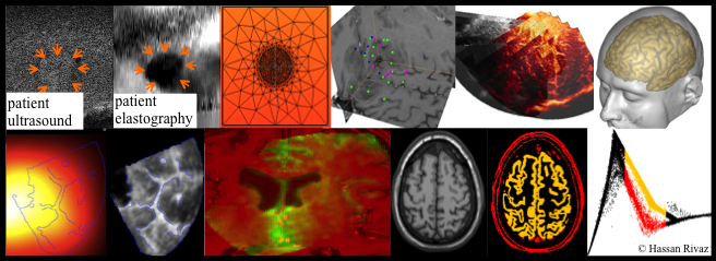
| Course description: | This course covers the principles and algorithms used in the processing and analysis of medical images. Topics include denoising, machine learning, image registration and similarity metrics. Image analysis methods on the most common medical imaging modalities (X-ray, MRI, CT, ultrasound) will be covered. Projects and assignments will provide students experience working with actual medical imaging data. |
| Prerequisites: | Undergraduate students: ELEC 364 or ELEC 342 Signals and Systems and a strong knowledge of linear algebra. |
| Credits: | Undergraduate course ELEC 444: 3 credits. Graduate course ELEC 6661: 4 credits. The graduate course requires carrying out a significantly more demanding project, which accounts for the extra credit. |
| Location & time: | FG B070, Thursdays 2:45 to 5:15 PM |
| Text Book: | Medical Image Processing, 2014, By Wolfgang Birkfellner, CRC Press, ISBN: 978-1466555570. |
| Reference Book: | Medical Image Processing, 2009, By Geoff Dougherty, Cambridge University Press, ISBN: 9780521860857. |
| Assignments: | Three assignments + one final project (in MATAB or another language based on student preference). Marker: Ms. Sepideh Khakzad, email: sepidehkhakzad AT gmail.com |
| POD | There are 3 PODs for this course: Ms. Noushin Jafarpisheh, Mr. Ali Tehrani and Mr. Sobhan Goudarzi. Please see the PDF file for POD timing and further details about the TAs. |
| Project: | All students (grad and ugrad) must be present in the last class (Week 13). The project due date is on Dec 6 at 11:59AM and should be emailed to Sepideh. |
| Project for undergrads: | Undergrads registered in ELEC 444 have an option of implementing a paper or doing a project based on the RANSAC algorithms. The students who choose the RANSAC project don't need to write a report and should email their code by Dec 6 at 11:59AM to the marker. |
| Grading: |
| Undergraduate (3 credits) | Graduate (4 credits) | |
| Assignments | 10 | 10 |
| Project | 20 | 40 |
| Midterm exam | 20 | 20 |
| Final exam | 50 | 30 |
| Assignments | Post date | Due date | Points |
| 1. Matrix and image manipulation | Wed Sept 7, 5PM | Wed Sept 21, 11:59PM | 1 point |
| 2. Detecting edges of brain MRI | Wed Sept 28, 5PM | Wed Oct 12, 11:59PM | 4.5 points |
| 3. Segmentation of brain MRI | Wed Oct 26, 9AM | Wed Nov 16, 11:59PM | 4.5 points |
| Outline: |
| Week | Topic |
| 1, Sep 8 | Logistics, introduction to X-ray, CT and nuclear imaging |
| 2, Sep 15 | Introduction to ultrasound and Magnetic Resonance (MR) imaging, images in Matlab |
| 3, Sep 22 | Ultrasound imaging and contrast agents |
| 4, Sep 29 | Convolution, aliasing in medical images |
| 5, Oct 6 | Denoising techniques in medical imaging, edge detection in medical images |
| 6, Oct 13 | Machine learning in medical imaging |
| Oct 20 | Midterm in class (mark your calendar) |
| 8, Oct 27 | Introduction to medical image segmentation, RANSAC and k-means in medical imaging |
| 9, Nov 3 | From linear filters to deep learning |
| 10, Nov 10 | Convolutional neural networks (aka CNN or ConvNet) |
| 11, Nov 17 | Registration of medical images, similarity metrics (sum of squared differences, normalized cross correlation, correlation ratio, joint entropy, mutual information) |
| 12, Nov 24 | AI in ultrasound |
| 13, Dec 1 | Student presentations |
| Databases: | These public databases of medical images may be useful for your project: 1) BrainWeb MRI link , 2) Ultrasound RF data link , 3) Ultrasound raw data link , 4) Fetal ultrasound head circumfrence: paper and data , 5A) Ultrasound data for segmentation provided by the paper below: Behboodi et al., RESECT-SEG: Open access annotations of intra-operative brain tumor ultrasound images, arXiv link, 5B) Ultrasound data for segmentation provided by the paper below: Yap, Moi Hoon, Gerard Pons, Joan Marti, Sergi Ganau, Melcior Sentis, Reyer Zwiggelaar, Adrian K. Davison, and Robert Marti. "Automated breast ultrasound lesions detection using convolutional neural networks." IEEE journal of biomedical and health informatics 22, no. 4 (2018): 1218-1226. 5C) paper Francesco Marzola, Nens van Alfen, Jonne Doorduin, Kristen M. Meiburger, Deep learning segmentation of transverse musculoskeletal ultrasound images for neuromuscular disease assessment, Computers in Biology and Medicine, 2021, 104623, ISSN 0010-4825 5D) Ultrasound data for segmentation and classification provided by the paper below: Al-Dhabyani, Walid, et al. "Dataset of breast ultrasound images." Data in brief 28 (2020): 104863. 5E) Ultrasound data (RF and B-mode) for segmentation and classification provided by the paper below: Piotrzkowska-Wroblewska, Hanna, et al. "Open access database of raw ultrasonic signals acquired from malignant and benign breast lesions." Medical physics 44.11 (2017): 6105-6109. 5F) TN3k Haifan Gong et al Multi-Task Learning For Thyroid Nodule Segmentation With Thyroid Region Prior, ISBI 2021 paper 5G) Unsupervised Contrastive Learning of Image Representations from Ultrasound Videos with Hard Negative Mining, Soumen Basu et al, MICCAI 2022 Gallbladder (GB) malignancy 6) Liver ultrasound: CLUST , 7) 2D Echocardiography, cardiac ultrasound. Paper: Leclerc et al, Deep Learning for Segmentation Using an Open Large-Scale Dataset in 2D Echocardiography, IEEE TMI 2019. the CAMUS Database, 8) Pediatric ultrasound brain segmentations. 1300 2D US scans for training and 329 for testing. A total of 1629 in vivo B-mode US images were obtained from 20 different subjects (age<1 years old): the US Database , the paper, 8) 3D MRI and ultrasound: the BITE Database, the RESECT Database And paper, the RESECT Database download , and MICCAI CuRIOUS Challenge 9) Ultrasound and CT of liver tumors. SYSU datasets. 10) 3D MRI of Brain: BRATS. 11) 3D MRI of Brain: WMH Segmentation. 12) 3D MRI of Brain: OASIS. 13) 3D MRI: Lumbar muscle and vertebral bodies segmentation of chemical shift encoding- based water-fat MRI: the reference database MyoSegmenTUM spine, 2019 14) Chest CT from dir-lab . 15) Chest CT from National Lung Screening Trial . 16a) Pancreas CT from TCIA Pancreas CT-82 . 16b) Kidney tumor CT, The kits19 challenge data: 300 kidney tumor cases with clinical context, CT semantic segmentations, and surgical outcomes, arXiv . 17) CT and MRI of several different organs Medical Decathlon . 18) CT, chest x-ray, digital pathology etc. Cancer Data Access System . 19) Liver CT LiTS - Liver Tumor Segmentation Challenge . 20) Abdominal CT IEEE TMI's DenseVNet Multi-organ Segmentation on Abdominal CT . 21) 50 abdomen CT scans from colorectal cancer chemotherapy from synapse, Beyond the Cranial Vault . 22) SCR database: Segmentation in Chest Radiographs, isi.uu.nl. 23) Inbreast Digital mamography link . 24) Curated Breast Imaging Subset of DDSM link . 25) Dataset of breast ultrasound images link . 26) A Large-Scale Database and a CNN Model for Attention-Based Glaucoma Detection, IEEE TMI 2020 link . 27) Cancer imaging archive link . 28) Hyperkvasir multi-class image and video dataset for gastrointestinal (GI) endoscopy link . 29) Kidney Ultrasound Data Set Data link , Paper link . |