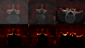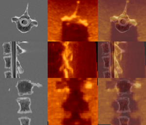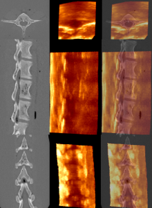


The repository includes three different datasets of corresponding CT and US images of vertebrae. In the first dataset, three human patients lumbar vertebrae are presented and the US images are simulated from the CT images. The second dataset includes corresponding CT and US images of a phantom made from post-mortem canine cervical and thoracic vertebrae. The third dataset presents the CT and US images of a phantom made from a post-mortem lamb lumbar vertebrae. For each of the two latter datasets, we provide 15 landmark pairs of matching structures in the CT and US images and performed fiducial registration to acquire a silver standard guidance of the registration.
Instructions for usage:
- All images are provided with NIFTI and MINC format.
- The first dataset can be used immediately after loading NIFTI or MINC images and the CT scans and simulated US images are aligned with a gold-standard ground-truth.
- The second and third datasets (the canine phantom and the lamb phantom) contain the CT scan, the intraoperative US, and the simulated US.
- For the CT scans and intraoperative US, 15 landmarks in MNI tag files are included so that they are aligned with a silver standard groundtruth and can also be used immediately after loading the NIFTI or MINC files.
- The CT scans and simulated US are aligned with a gold standard ground-truth.
Please cite this paper if you use this data:
N Masoumi, C. Belasso, M Ahmad, H Benali, Y Xiao, H. Rivaz, Multi-modal 3D ultrasound and CT in image-guided spinal surgery: public database and new registration algorithms, Springer IJCARS, 2021

